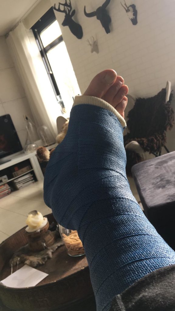
Here you see a picture of the unfortunate patient with his right lower leg in plaster. In the meantime, the plaster has been removed and the patient is again walking carefully without aids. Now 7 weeks have passed since the trauma. Dynamic ultrasound examination showed that the butt ends do not give way; we may assume that very dosed loading is now possible. The prognosis is good if there is no relapse trauma. Caution is therefore still required, even in everyday matters such as putting on and taking off socks! So far so good, you might say…
The question remains whether the diagnostics are ‘lege artis’. Here I speak for myself. As far as the Thompson test is concerned, with its sensitivity and specificity above 0.9, it scores very high. However, even with this test, the danger of a false negative score (no AP rupture) is lurking, because when squeezing the calf musculature, other flexors than the AP plantar flexion can cause it. Active plantar flexion is not impossible even with full thickness rupture of the AP if the plantar muscle is intact!
For clinical research the following is important:
- Thompson test + ; 2. loss of strength at plantar flexion; 3. a ‘del’ at the level of the tendon tear; 4. a hematoma (usually distal from the tear).
If the above refers to a ‘tear’, we still do not know for sure whether this is a tear in the AP or in a gastrocnemius tendon, or whether we are dealing with a muscle tear? The two ruptures as I saw in this case are – if so – not observed without imaging diagnostics.
Is it necessary to know exactly where the lesion is situated if the treatment is: plaster! I think so; a standard echo shows if and how much retraction there is from the butt ends, if the paratenon is intact and if there is fat from the Kagerian fat body between the butt ends. With the above, the prognosis is not very favourable and surgery should be considered if the strain on the tendon is an issue. The patient in this case received the right therapy, fortunately. There was some luck because no one had any idea in advance of the imaging of the location of the tendon.
At Voorhorst