Pictures: EchoProcedures/Voorhorst
Where do you find a medial plica in your knee?
In my previous article about the M. articularis genus, the medial plica in the knee was mentioned. This muscle and the plica have a lot to do with each other. I’ll write more about that later. With these images, I looked for a possible plica in my own knee. A plica is a fold in the synovial capsule of the knee and is considered a remnant from the embryonic development. Many people have a plica on top of the knee, on the inside or on the underside of the kneecap. Most plicae are asymptomatic, we have no complaints. Sometimes, however, they become inflamed by frequent knee bending or trauma, such as a kick during football or bumping against the edge of the table. Then a plica can become irritated with all its unpleasant consequences. It is still useful that – as caregivers – we have the plica syndrome in our minds as the cause of (medial) anterior knee pain.
On picture B the place where – in the case of a thickened plica – you can feel an edge that moves back and forth under your finger (if the knee is held in the stretched position).
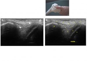
Left photo A is the same as photo B.
In A: the white arrows point to the synovial membrane. In B: P: patella (kneecap); f: femur (upper leg); c: cartilage of the upper leg; f: fat body in the joint; ** the joint capsule (consists of two layers, the fibrous capsule and the synovial capsule); rrrr: retinaculum of the patella.
At Voorhorst