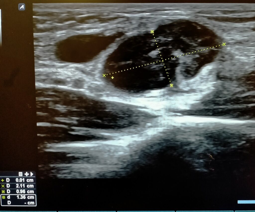
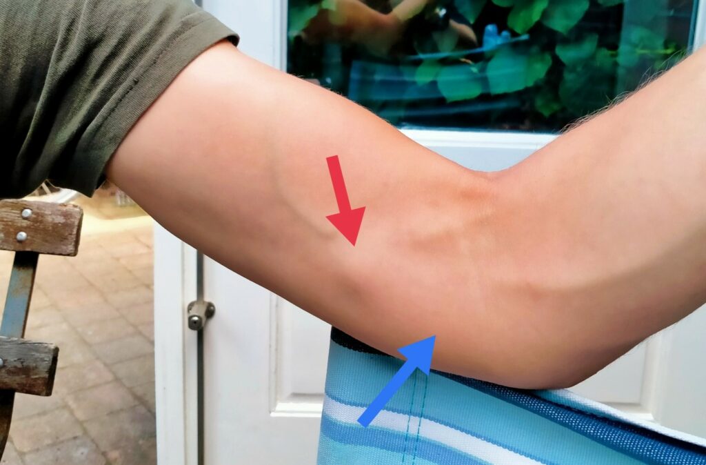
Sonography of an enlarged hyperintense epitrochlear lymphnode (left arm) in a
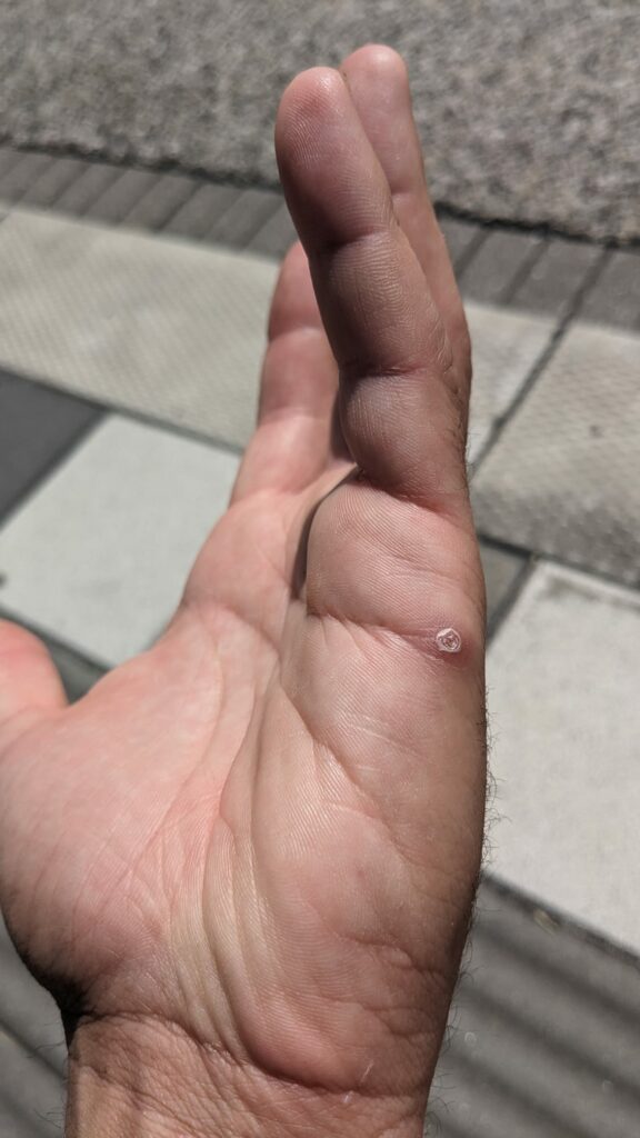
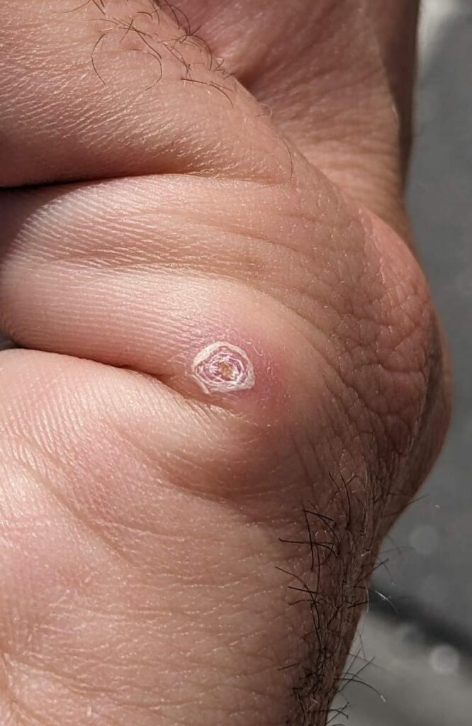
33 year old male; and an ipsilateral ulcerated papula, suspected to be the port d’entree for
Cat Scratch Disease. Serologically not confirmed – at this moment – for bartonella henselae.
(photo: At Voorhorst)
(photo: At Voorhorst)

(photo: At Voorhorst)
(photo: At Voorhorst)
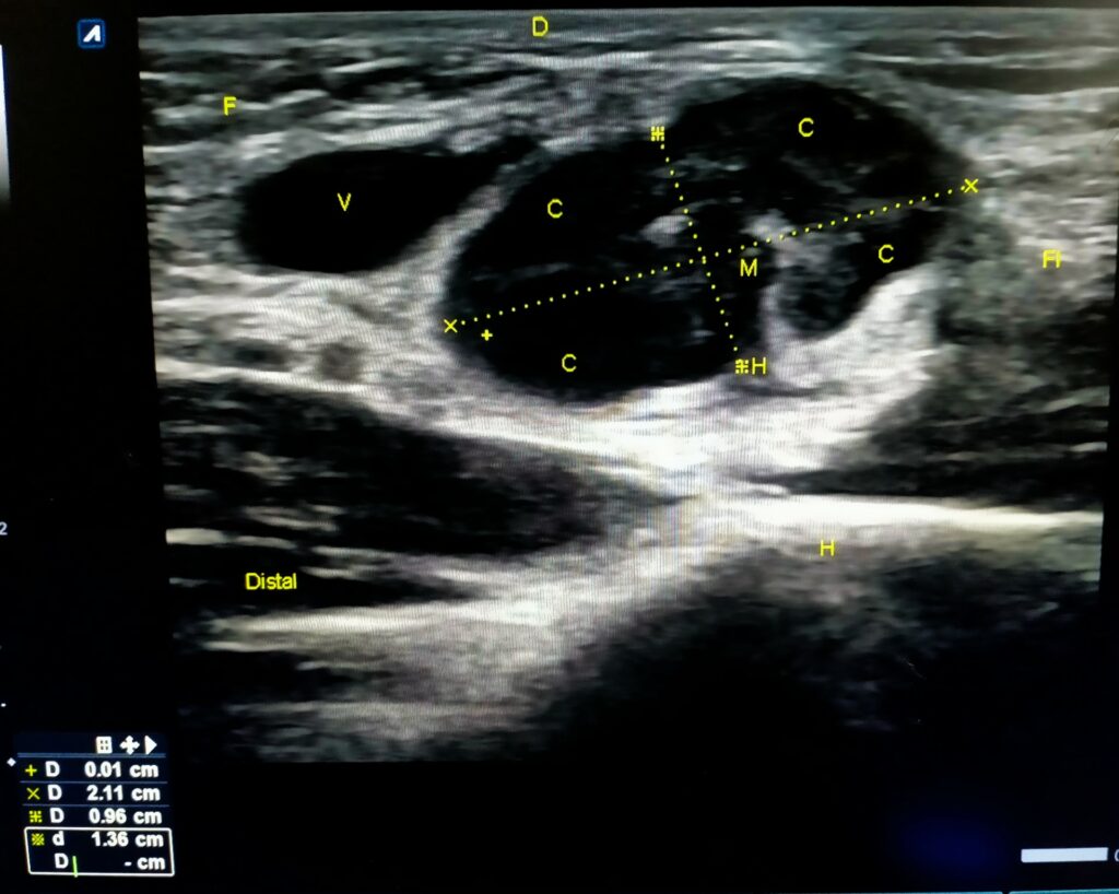
H; hilum/hilus; C: cortex; M: medulla
(photo; At Voorhorst)
Thanks for your support Arne Voorhorst!